
Fluorescent Confocal Microscopy
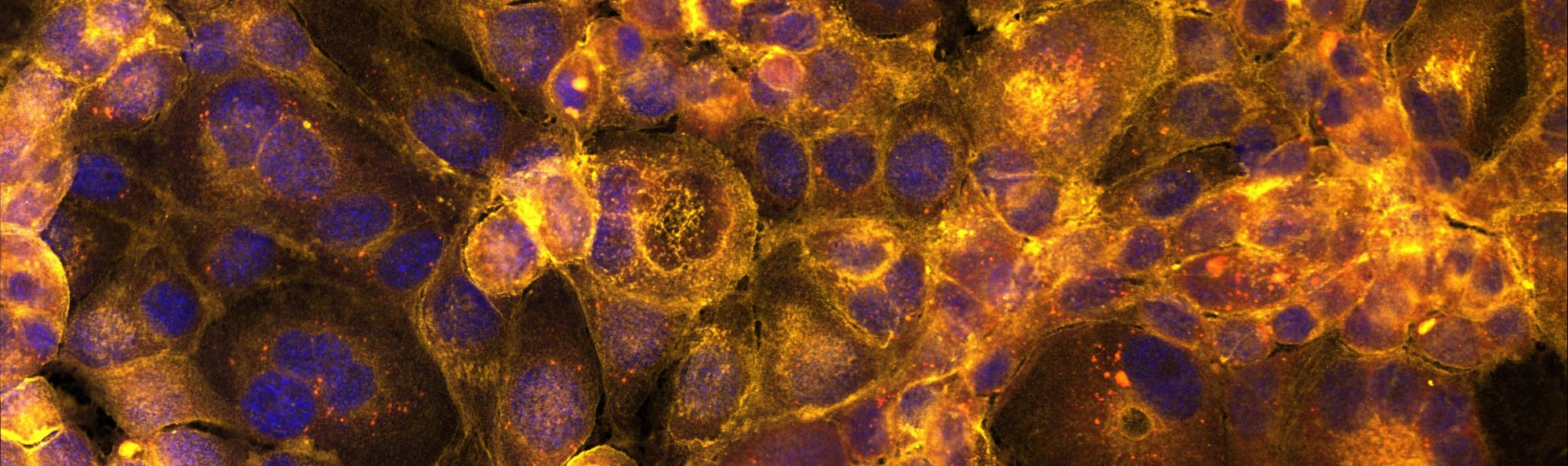
2024 Scholars Week Scientific Image Competition People's Choice Winner: "Galactic Patterns of Hepatocytes" by Kaya Wright
About
Fluorescent laser-scanning confocal microscopy (LSCM) is an optical imaging technique that can be broken down into three main components: fluorescence, laser-scanning & confocal. Fluorescent microscopy detects the presence and localization of fluorescent molecules in a sample. This is achieved through the scanning of a laser tuned to a specific wavelength of light that excites fluorescent molecules in the sample. This excitation results in emission of photons that are then received by the detectors and translated to pixels of the raster image. Confocal microscopy reduces blur and modestly increases image resolution by blocking out out-of-focus light. This enables the creation of 3D images of your sample by layering images (also called optical slices) in a 'z-stack'. This instrument was purchased with funds from the National Science Foundation Major Research Infrastructure Program (NSF DBI 2019228)
Located BH 016
Primary Contact
Alyssa Tsukada
she/her
tsukada2@wwu.edu
(360) 650-7577
ES 508d
How to Get Started
Check out our New Users page!
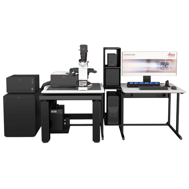
Leica Stellaris 8 Laser Confocal Fluorescent Microscope
Primary Uses:
Optical sectioning confocal, FLIM, FCCS, Super-resolution, FRAP, DIC
Objectives:
10X NA 0.4, 20X NA 0.75, 25X NA 0.95 NA (water), 40X NA 1.4 (oil), 63X NA 1.4 (oil), 63X NA 1.2 (water)
Detectors:
5 HyD detectors with spectrometer for spectral scanning from 410-850 nm
Additional Assets
Computer – HP, Intel Xeon Gold w/ 8 cores, 192 GB RAM, NVIDIA GPU w/ 24 GB RAM
Software – LasX with Tau-sense, Phasors, Lightning, 3D Visualization
Stand – Leica DMi8 automated microscope
Stage – Encoded, closed loop automated stage with autofocus and Piezo Z axis
Illumination – 80 MHz pulsed white light laser tunable in 1 nm increments from 440-790 nm with 8 lines available simultaneously; 405 nm continuous laser
Confocal – Stellaris8 resonance scanning confocal scan head
Super-resolution – Lightning deconvolution based super-resolution to 150 nm
Time-correlated photon counting – Enables fluorescence lifetime measurements and fluorescence cross correlation spectroscopy
Sample Preparation
More resources coming soon!
Educational Content
Intro to Fluorescent Confocal Microscopy
Confocal Microscopy and 3D Recontruction
Intro to LasX Software
Published Data
1. Lee, S.R., Pollard, D.A., Galati, D.F., Kelly, M.L., Miller, B., Mong, C., Morris, M.N., Roberts-Nygren, K., Kapler, G.M., Zinkgraf, M., et al. (2021). Disruption of a ∼23–24 nucleotide small RNA pathway elevates DNA damage responses in Tetrahymena thermophila. MBoC 32, 1335–1346. 10.1091/mbc.E20-10-0631.
2. Novis, P.M., Dhami, M., Podolyan, A., Matsumoto, M., and Kodner, R. (2023). The austral biflagellate Chloromonas rubroleosa (Chlorophyceae) is the closest relative of the unusual quadriflagellate genus Chlainomonas, both found in snow. Journal of Phycology 59, 342–355. 10.1111/jpy.13318.
3. Galati, D.F., and Asai, D.J. (2023). Immunofluorescence Microscopy. Current Protocols 3, e842. 10.1002/cpz1.842.
Submission Form
If you have utilized any SciTech resources for research that has been published or presented at conferences please fill out this form.